Sunday, December 9, 2007
New research on the blind is revealing the brain's ability to adapt -- and may lead to new therapies for everything from strokes to chronic pain
Courtesy By Cara FeinbergSource : Boston Globe
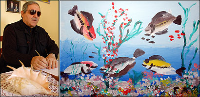
Esref Armagan is a 52-year-old Turkish painter who has been blind in both eyes since the day he was born. He has never seen a coffee cup, a toothbrush, an elephant, or a tree-lined street, but he can draw them each, from any perspective, with or without shadows depending on the time of day. His portrait of President Clinton, which he painted from an embossed photograph, looks, well, like Clinton -- complete with grey hair and bulbous nose -- and though Armagan has never had an art lesson, the streets he paints stretch into the distance as converging parallel lines.
For years, Armagan has been a phenomenon in the art world, displaying his work in museums around the globe. But it was not until two summers ago, when he traveled to Boston, that scientists were able to study precisely how he generates such images. Their hope was that he might teach them something about neural ''plasticity"--the brain's ability to reorganize its functions based on new information and experiences. If Armagan had never seen with his eyes, how had his brain adapted to give him visual representations of the world, and more importantly, what could it reveal about brain adaptation in general?
In July of 2004, at the Center for Noninvasive Brain Stimulation at Beth Israel Deaconess Hospital in Boston, Armagan agreed to have his brain imaged in a magnetic resonance imaging (MRI) machine while he drew with a pencil on a sheet of paper. He explored a set of objects by touch -- a coffee cup, a toy elephant, a toothbrush -- and then was told to imagine them and draw them all from memory. Each time, his drawings hit the mark.
''What we saw in the scan was quite amazing," says Dr. Alvaro Pascual -Leone, an associate professor of neurology at Harvard Medical School and director of the center. He and two colleagues in Beth Israel Deaconess's neurology department, Amir Amedi , PhD, and Dr. Lotfi Merabet , conducted a series of scans, each time challenging Armagan with more complex tasks. '' Esref's visual cortex lit up during the drawing tasks as if he were actually seeing," says Pascual -Leone. ''His scan, to the untrained eye, might look like the brain of a sighted person."
Armagan presented a unique learning opportunity for the scientists at Beth Israel Deaconess. Pascual -Leone and his colleagues had access to a blind person able to render -- pictorially -- what his mind's eye had captured. But more importantly, they now had the technology to look at his brain while he rendered it, and to glimpse how his visual cortex functioned after 52 years without vision.
For centuries, scientists held that the brain was a fixed entity, that it was hard-wired for each individual function, and incapable of reorganizing after injury. In the late 1850s, the French neurosurgeon Paul Broca was the first to argue that language was associated with a specific part of the brain, and other investigators soon followed suit: The visual cortex at the back of the brain, they hypothesized, processed only vision, the somatosensory cortex in the mid-brain processed only pain, vibration, and touch, the auditory cortex on the sides of the brain existed solely to process sound.
In the last half-century, however, new technology and cutting-edge experiments like those of Pascual -Leone and his colleagues, have exploded that dogma, revealing not only that the brain does in fact reorganize and adapt, it does so all the time. ''What we saw in Esref ," Pascual -Leone explains, ''was that he was using his visual cortex. It wasn't lying dormant. It hadn't shrunk or disappeared. Instead, it was recruited by other senses."
The brain, as work like Pascual -Leone's is revealing, is a lot more resourceful than we ever knew it was.
How a Blind Person's Brain Can "See"
Dr. Pascual -Leone has been studying the brain for three decades, examining its capacity to establish new neural connections, how to use the connections that exist, and how to harness them to create better rehabilitation strategies after trauma or sickness.
Pascual -Leone's patients and study subjects range from normal-functioning adults with special gifts like Armagan , to those with a range of neurological deficits, from sensory loss, to strokes, to chronic pain, to medically-resistant depression.
The blind, Pascual -Leone explains, provide an excellent opportunity to study brain plasticity. ''A large part of our brains is devoted to vision-some estimate more than half," he says. ''The question we are asking is what happens to that part of the brain when there is no input from the eyes?"
Over the past 10 years, Pascual -Leone and several other scientists, including his colleague Amir Amedi , have conducted experiments examining the brain's role in sensory perception, and much of their work has been with blind subjects. Using neural scans and transcranial magnetic stimulation ( TMS )-a technique in which a noninvasive handheld device is used to stimulate or temporarily interfere with targeted brain functions-several studies have found activation in blind subjects' visual cortices, despite the fact they cannot see.
In an early study Pascual -Leone coauthored with Dr. Leonardo Cohen at the National Institutes of Health, results showed that during Braille-reading tasks, blind subjects' visual cortices lit up like lamps. But the mere fact that there was activity in that section, he pointed out, did not necessarily prove it was vital to that function. That, he said, is where TMS comes in.
''If you use TMS to temporarily interfere with the visual cortex during certain tactile tasks, like reading Braille, you'll find that early-blind subjects suddenly have trouble performing them," says Amedi , whose own independent studies have revealed similar results during language-based tasks. In the blind, unlike the sighted, the TMS interference, researchers believe, shows that the visual cortex is engaged-and in fact required-for certain nonvisual tasks.
So if, as scientists' findings suggest, the visual cortex need not be devoted solely to sight, how does the brain adapt after injury or new environmental influences? Does the brain forge new connections that did not exist before, or are the connections already there lying dormant, pressed into service by the circumstances?
Pascual -Leone's current work with his colleagues at Beth Israel Deaconess aims to answer those questions. For the past few years, they have been studying sighted subjects who volunteer to be blindfolded for five days and learn certain nonvisual tasks, including rudimentary Braille. In every case, before subjects donned the blindfold,functional MRI ( fMRI ) scans revealed little activity in their visual cortices during tactile tasks. After the subjects wore the blindfolds for two days, however, the scans showed bright patches of activity in the visual brain when the subjects used their fingers for tactile or Braille-reading tasks. By day five, the visual cortex glowed steadily during these same tasks. Yet two hours after the blindfolds were removed and the subjects' eyes had readjusted, scans of the visual area of their brains were as dark as they'd been on day one. Once the blindfolds were removed, touching, handling objects, and Braille-reading no longer activated ''sight" in the seeing.
The cortical adaptations that occur in the blindfold studies appear -- and disappear -- too quickly for any new nerve connections to grow, Pascual -Leone believes. He compares the adaptive pathways in the brain to detours after road blocks; building a new street takes a long time, he explains, but if there are other existing surrounding roads, they can be used right away. These immediate neurological detours reveal the brain's capacity to adapt in response to environmental factors, but it is sustained sight loss that will more likely result in lasting adaptations, he says.
Over time, if the brain continues to follow the detour routes, he believes, it starts to modify them to make them better, and might even make new structural connections. The fact that change occurred so immediately in the blindfolded subjects, he says, indicates that the visual cortex may inherently possess the machinery necessary to process nonvisual information.
Pascual -Leone and his colleagues believe that humans work with a reserve of existing connections dictated by their specific genetic make-up that, depending on their use, will become masked or unmasked by the individual's circumstances. ''What Esref and the blindfold studies show us," he says, is that lacking sight, the brain draws on information from the other senses. ''Even in the absence of vision," he says, ''the visual cortex is involved in creating images." In other words, the work of Pascual -Leone and others suggests that the brain has many additional capacities it can call on in a pinch.
The Birth of Brain Therapy
As both a physician and a researcher, Pascual -Leone aims to put his findings from his studies of the blind to use in developing rehabilitative therapies for other types of conditions. But his lab is not alone in its development of new treatments.
Other breakthrough therapies have arisen for strokes, autism, schizophrenia, spinal cord injuries, epilepsy, chronic pain, and many other previously ''untreatable" conditions. At the University of California, San Francisco, one neuroscientist has developed a computer program to teach language skills to dyslexic children through what is called, ''neural retraining." A professor in the department of psychology at the University of Alabama has used these developments to help stroke victims restore movement in their limbs. Two scientists at the University of Rochester have found that playing action video games can enhance a range of visual attention skills.
Yet despite the dozens of medical therapies that have been developed as a result of breakthroughs in thinking about brain plasticity, says Pascual -Leone, in both our scientific understanding of these mechanisms, and our ability to apply them clinically, we are still at the starting gate.
For researchers studying the brain, the next steps lie in learning enough about plasticity to harness it for individual needs. Through their work with the blind, Pascual -Leone and his colleagues hope to learn more about how visual images can be processed nonvisually in the brain-both for what it will tell them generally about how the brain works, and how, specifically, they might help the brain to work better for the newly blind or those who regain sight.
Subjects like Esref Armagan , says Pascual -Leone, help jump-start that process. ''We can never know what types of images were actually being created in Armagan's brain," he says. ''But we know now that when he draws those images, we can understand them visually without a doubt." This makes it seem as if he is seeing, says Pascual -Leone. ''And when we looked at his brain, we could see how." Edited and Compiled by: Ashutosh Pandey/ Anju.
Posted by GEET Education
|